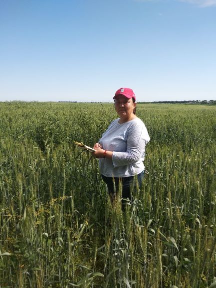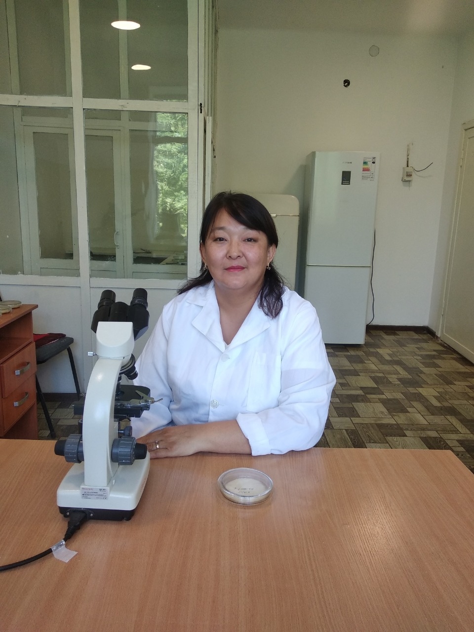
Scientists of Zh. Zhiyembayev LLP "Kazakh research institute for plant protection and quarantine” talked about the varieties of diseases of cereals with seed infection and methods for their diagnosis. During the consultation for farmers, Kopirova Gulsim Islamovna, a researcher at the department of grain, oilseeds and legumes, shared her scientific observations and developments on this topic.
G. Kopirova noted that every year due to diseases farms do not get on average up to 30% of the possible grain harvest. As a result, not only the quantity of the harvest is reduced, but also the quality. About 60% of phytopathogens are transmitted through seeds. Often there are cases of latent infection, without external signs, when the infection is detected only during phyto-examination. Contamination of microflora of seed material can occur at different stages: during the growing season, during harvesting, especially in the presence of high humidity, during threshing or after processing after harvesting; during storage of the crop, in case of violation of the storage regime, as well as when collecting seeds in storage areas with high humidity.
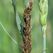
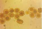
The microflora found in the seeds is divided into saprophytic (penicillas, aspergillus, mucor and alternaria, etc.) and pathogenic (fungi causing diseases such as smut, root rot, fusarium wilt, septoria, etc.).
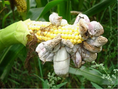
The following types of cereal diseases are transmitted by seeds: covered smut, dust brand, hag and dwarf smut of wheat, helminthosporious, fusarium root rot, tungaltera of cereal crops, septoria, helminthosporious spotting, fusarium wilt, various bacterioses and viral diseases of wheat. Stone, dusty and false dusty smut of barley, dark brown, banded spotting, root rot of barley. Covered and black smut and helminthosporious spotting of oats. Covered smut millet. Bubble and dusty smut of corn. Pyriculariosis, helminthosporiosis and fusarium wilting of rice.
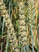
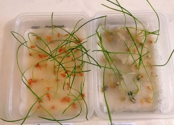
Contamination of cereal seeds with a complex of pathogens can be established in laboratories when determining their sowing qualities with additional tests for contamination of their covered smut and smut-covered centrifugation and microscopy, and infection with septoria and helminthosporiasis by germinating them on paper rolls.
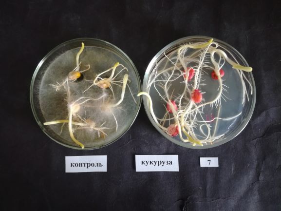
An external inspection is carried out simultaneously with the analysis of the purity of the seed material, while the presence of impurities of smut smut sacs and ergot horns in the grain is established. Contamination of cereal seeds with glume mould of wheat, fusarium cladosporiosis, etc. is determined on a sample isolated from the original sample. Seeds are scattered on glass with a thin layer and divided by a ruler into 4 triangles. 100 grains were selected from each and the degree of seed damage by the black embryo and the darkening of the films on a scale of A.G. Tropova was visually established.
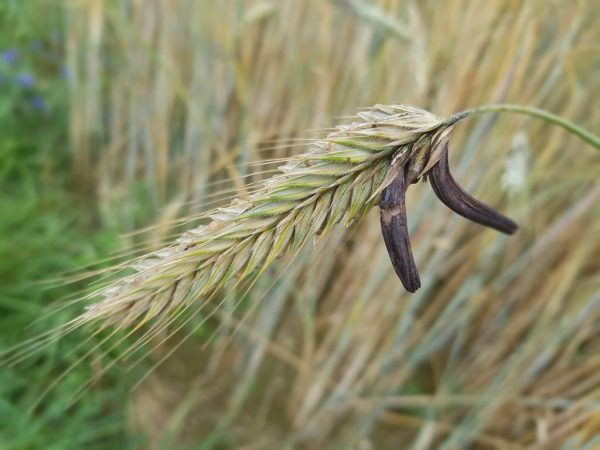
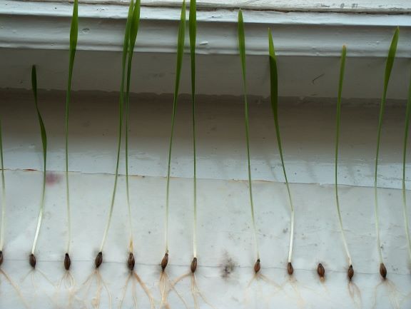
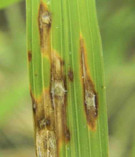
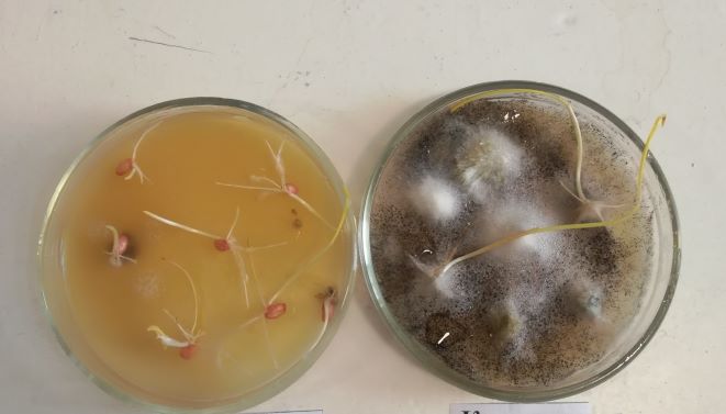
Methods of washing and centrifugation establish seed clogging with teliospores of external smut species, as well as conidia of fungi helminthosporium, fusarium, alternaria and septoria. For this purpose, 2 tubes of 100 grains are taken from the average sample, they are placed in clean tubes, 10 ml of water are poured and shaken for 10 minutes. After this, it is poured into clean special tubes and centrifuged for 5 minutes at a speed of 1000-1500 rpm. After the water is drained from the test tube, the sediment is stirred up, a drop is applied to a glass slide and viewed under a microscope. To determine the infection of seeds with fungal and bacterial infections, the method of wet chambers and sowing on special nutrient media are used. For this purpose, the grains are sterilized in alcohol for 2-3 minutes or with 0.5% KMnO4, 10-20 minutes, then washed with sterile water and laid out on moistened filter paper or on a nutrient medium and exposed in a thermostat for 3-5 days at a temperature of 20 -25 C. To determine the infection of seeds with various pathogens, they are germinated on paper rolls until 2-3 leaves appear. The imprint method is used instead of centrifugation to determine the contamination surfaces of cereal seeds with smut fungi. Each seed is wrapped with a piece of transparent adhesive tape 1 cm in size, tightly pressing it over the entire surface of the seed. Then, using tweezers, the tape is peeled off and placed on a microscope slide to identify the pathogen and count the spores. Counting spores is carried out in 10 fields of the microscope in contact with the seed parts of a piece of tape and set the arithmetic mean number of spores in one field of view of the microscope.
